MRI & X-Ray
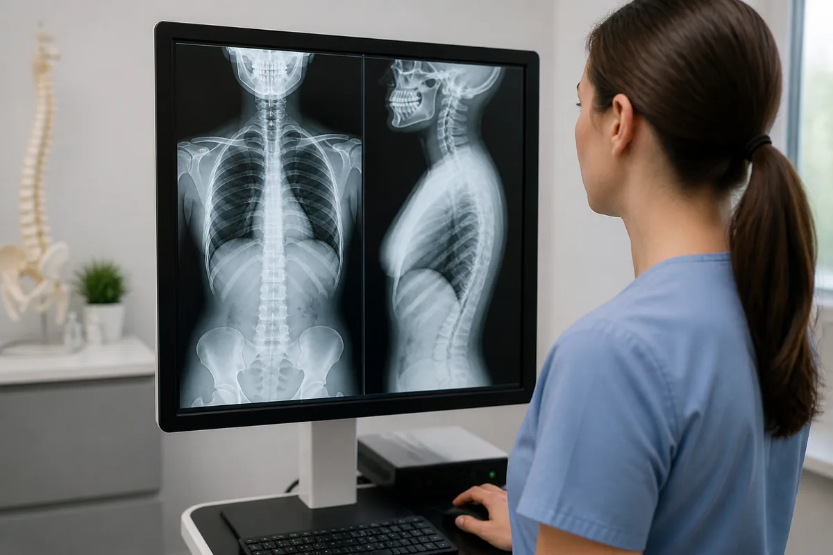
MRI & X-Ray
MRI and X-ray imaging give a clear picture of what is happening inside the body, from bone fractures to disc and soft-tissue problems. Having these studies on site means your imaging and your physical therapy plan stay connected, without extra trips to an outside facility.
What Each Study Shows
- X-Ray — a quick image used to check bones and joints, often the first step after an injury or fall.
- MRI — detailed imaging of soft tissue, discs, nerves, and joints, useful when symptoms point to a herniated disc, nerve pain, or a soft-tissue injury.
How Imaging Supports Your Care
Once imaging is complete, a radiologist's report is sent to your physician or therapist so it can directly inform your treatment plan. This is especially useful for patients dealing with back or neck pain, sports injuries, post-surgical recovery, or nerve-related symptoms, where seeing the underlying structure helps guide the right approach.
Why On-Site Imaging Helps
- Convenience — imaging and therapy happen under the same roof, reducing the back-and-forth between separate offices.
- Coordinated Care — your treatment team can review results and adjust your plan without waiting on outside records to arrive.
- Clear Communication — reports are shared with your physician or therapist, keeping everyone on the same page about your recovery.
This content is for general information only and isn't medical advice. Check with your provider or doctor before you start a new exercise or treatment. If you have a medical emergency, call 911.